And the winners are…

2025 WINNER IN THE MOST STUNNING CATEGORY
“Guardians in Conversation” by Shikha Srivastava, university of louisville. imaging facility: MNTC huson lab (uofL). Description: Two neutrophils, the body’s first defenders, connect with each other as if quietly communicating during an infection. This small interaction shows how they work together, turning a tiny moment into a powerful display of teamwork and defense.
2025 WINNER IN THE MOST UNIQUE CAPABILITY CATEGORY
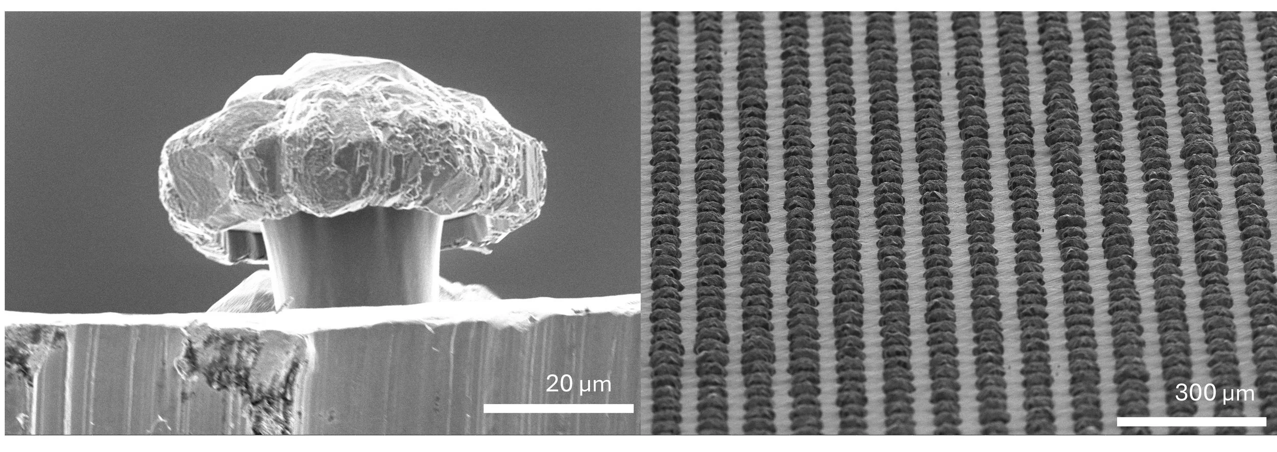
“Micro-Mushroom array” by hyogyun roh, conn center, University of Louisville, EMC. Imaging facility: MNTC Huson lab. DESCRIPTION: fabricated "doubly-re-Entrant structured Cu mushroom array”. This material was used to create a superhydrophobic surface. The images show the doubly re-entrant structure of micromushrooms in the underside view (left) and uniform mushroom array in the top view (right).

2025 winner in the most whimsical category
“Love owl” by Matthew Mulvehill, Conn Center for Renewable Energy Research (uOFL). imaging facility: MNTC huson lab (uofL). description: A scanning electron microscope (SEM) image of the left-over stump of a titanium dioxide nano-rod coated silicon micro-pillar that broke off from the substrate. The dark shadow around the owl’s eyes is silicon, the unique shape being an undesired artifact of a dry reactive ion etch (DRIE) during pillar formation. The silicon is coated with a thin layer of fluorine doped tin oxide followed by the titanium dioxide nano-rods that blanket the entire surface. The micro-pillar that was previously attached to the owl’s face will be used to sustainably generate hydrogen from only water and sunlight.
Competing in the Most Unique Capability Category

“Micro-Mushroom array” by hyogyun roh, conn center, University of Louisville, EMC. Imaging facility: MNTC Huson lab. DESCRIPTION: fabricated "doubly-re-Entrant structured Cu mushroom array”. This material was used to create a superhydrophobic surface. The images show the doubly re-entrant structure of micromushrooms in the underside view (left) and uniform mushroom array in the top view (right).

“Corrosion of Copper Metal Strips” by Waliul Islam Khan, Department of Chemistry, University of Louisville. Imaging facility: MNTC Huson Lab (UofL). Description: corrosion of copper metal strip in 2 weight% gel prepared in 1 weight% aqueous sodium chloride solution.

“Copper Hills” by Fahad Bin Halim, chemistry department, university of louisville. imaging facility: mntc huson lab (uofl). description: Tool – Apreo C LoVac FESEM ( Thermo Fisher ) Uniform Copper (Cu) hill-like formations were observed when Cu was electrodeposited at Bulk deposition conditions on clean Bare Au electrodes. These high-rough 3D Cu nanostructured surface on Au electrode boosts active surface area and local fields for use in electrocatalysis, non-enzymatic sensing and SERS/ SEIRA substrate. The height and roughness of such Cu hills can be controlled by deposition parameters allowing formation of user-defined templates.

“flooded forest” by Matthew Mulvehill, Conn Center for Renewable Energy Research (uofL). imaging facility: mntc huson lab (uofl). Description: A scanning electron microscope (SEM) image of a forest of titanium dioxide nano-rod coated silicon micro-pillars. The titanium dioxide nano-rods are coated in a thin layer of carbon via sputtering, resulting in a more bloated appearance of the nano-rods. The bases of the pillars are submerged in a polymer that serves as protection for the carbon that lies underneath from plasma etching to clean the tops and sides of the pillars. The cleaned micro-pillars are then used in photo-electrochemistry experiments to learn how well the micro-pillars absorb light and use that energy to drive chemical reactions.
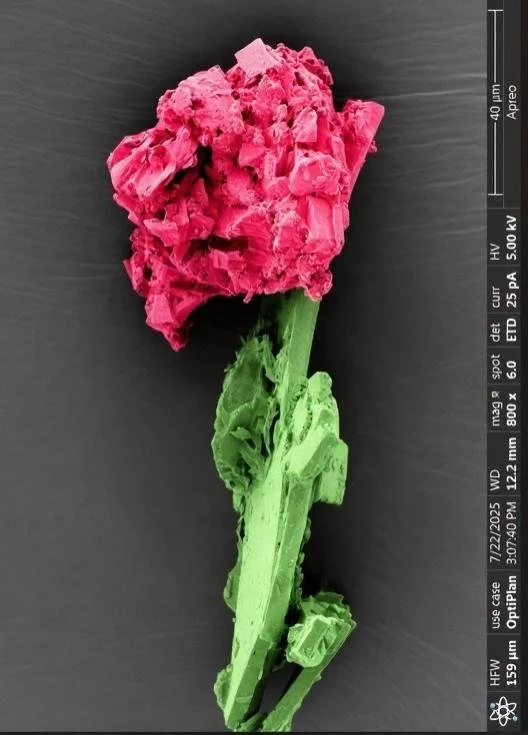
“Nano Rose on a Stem” by Sharmin Akter, university of louisville. imaging facilty: mntc huson lab (uofl). Description: This SEM micrograph, captured at the Micro/Nano Technology Center (MNTC) of the University of Louisville, shows a perovskite powder particle. The sample was prepared as part of the precursor material used for coating the carbon electrode layer in carbon-based perovskite solar cells.

“un-named” by julia aebersold, uofl mntc. imaging facility: mntc bioEM lab (uofl). Description: TEM images of Treponema Denticola. This spiral-shaped bacteria is found in the mouth and known for its ability to invade tissues and cause inflammation and bone loss. To help prevent the issues with these critters brush your teeth twice a day and floss daily.
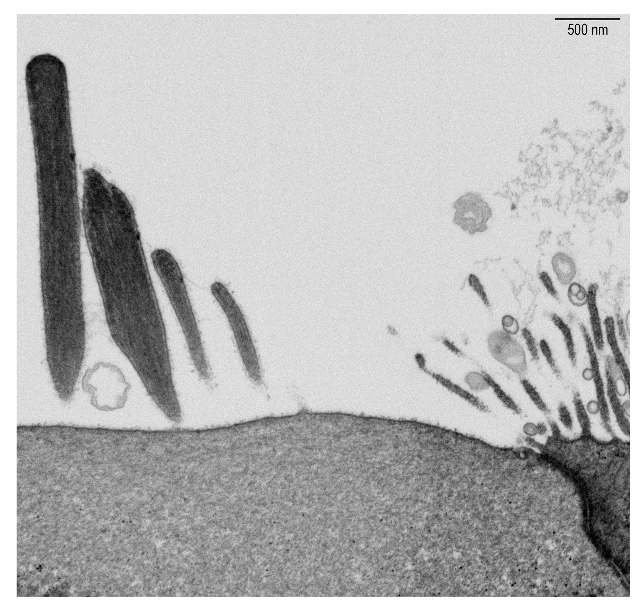
“One of Two of a kind” by Abigail Dragich, University of kentucky. co-author: Gregory Frolenkov. Imaging facility:EMC (UK). Description: Photo description: Auditory hair cells are mechanosensitive cells in the cochlea responsible for detecting sound so we can hear. Each cell has a hair bundle structure made of individual stereocilia projections with specific ultrastructural features. Here, with 20nm FIB serial sectioning, we capture a cross-section through four adjacent stereocilia showing all three tip links interconnecting them and three electron dense upper tip link densities. This is only the second photo ever to show all these features together; the other image comes from a classical transmission electron microscopy section of ~50nm. Image was taken in the UK EMC with FEI Helios Nanolab 660.

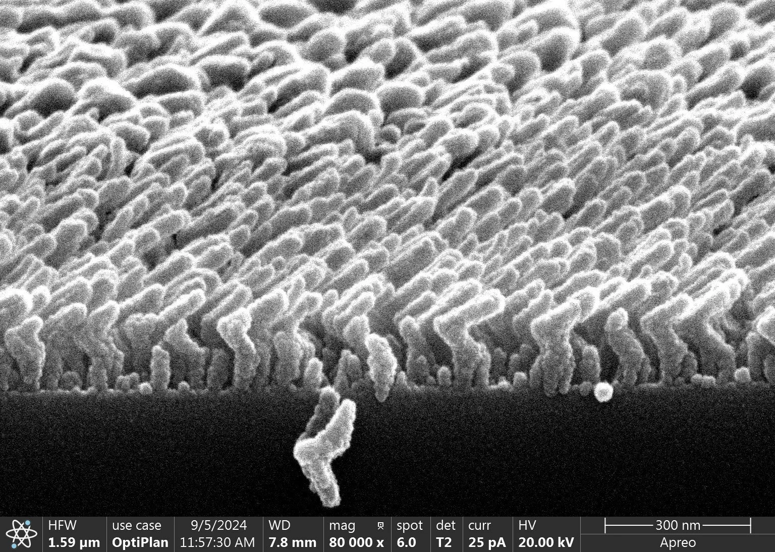
“Echoes of Structure” by Ana Isabel Lopez-Porras, university of kentucky. imaging facility: EMC (UK). Description: High-resolution image acquired using UKy Focused Ion Beam-Scanning Electron Microscope (FIB-SEM) system, showing Prominent, elongated surface projections called stereocilia, on the apical surface of a sensory hair cell. The dense actin core of each stereocilium is visible, projecting from the upper hair cell membrane. The image captures the internal organization of the cell, including mitochondria and other organelles beneath the apical membrane.
“Field of Nanoboomerangs” by chuang Qu, U. of Colorado,.imaging facility: MNTC huson lab (uofL). Description: This SEM image features tiny boomerangs (sub 100 nm critical dimensions), appearing as if they are sprouting from the substrate field. The Germanium boomerangs are grown by glancing angle deposition (GLAD) using e-beam evaporator at MNTC at UofL cleanroom, and imaged at Huson lab at UofL.
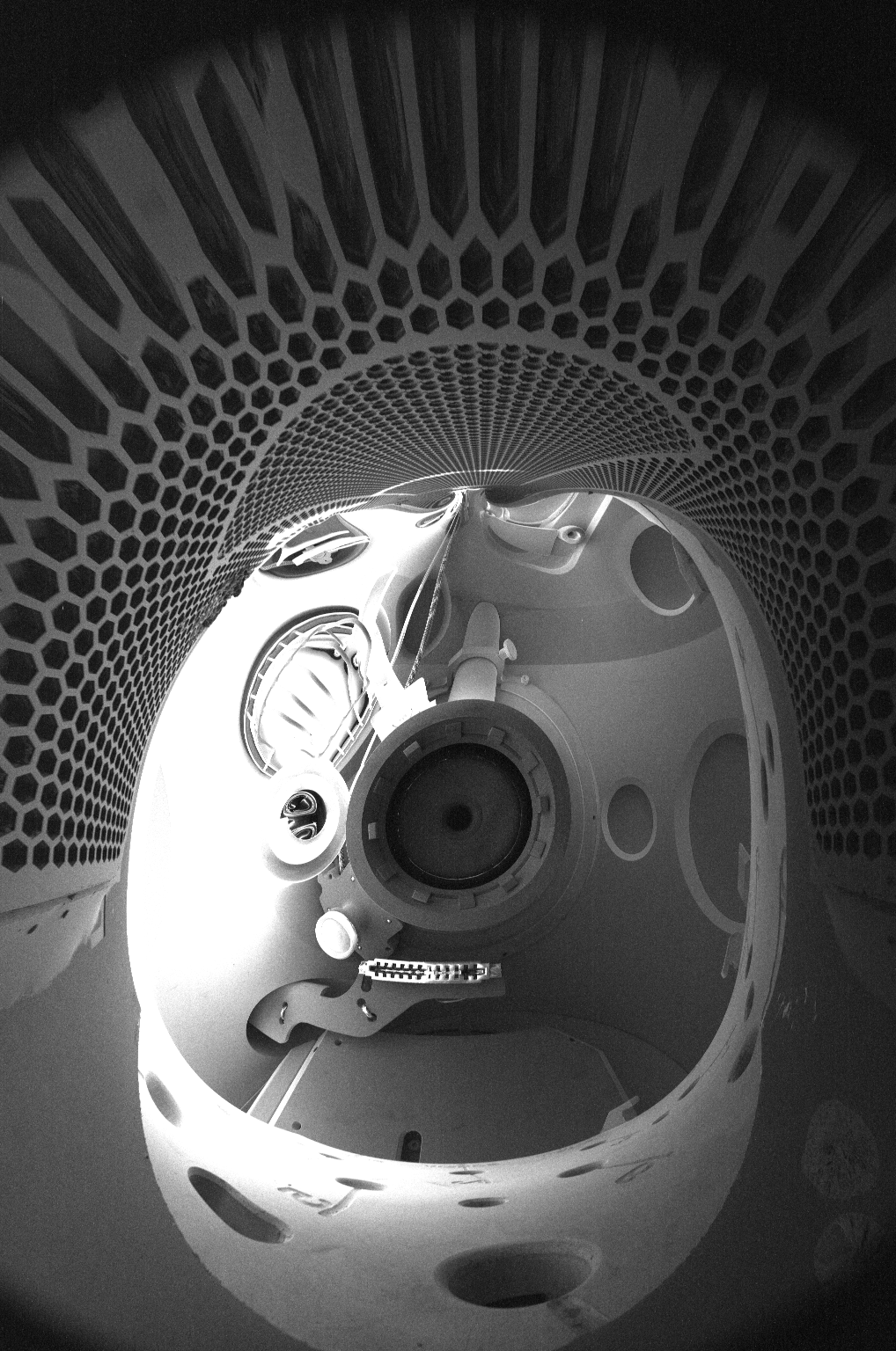
“un-named” by michael martin, uofl mntc. Co-author: Belinda Petri, Department of Biochemistry and Molecular Genetics, University of Louisville School of Medicine. imaging facility: mntc huson lab (uofl). description: This is an electrostatic reflection from a polymer sheet that makes up a Nanopore RNA sequencing device. In the image we see both the hexagons on the device's surface and the pole piece/detector that is taking the image.
Competing in the Most Whimsical Category

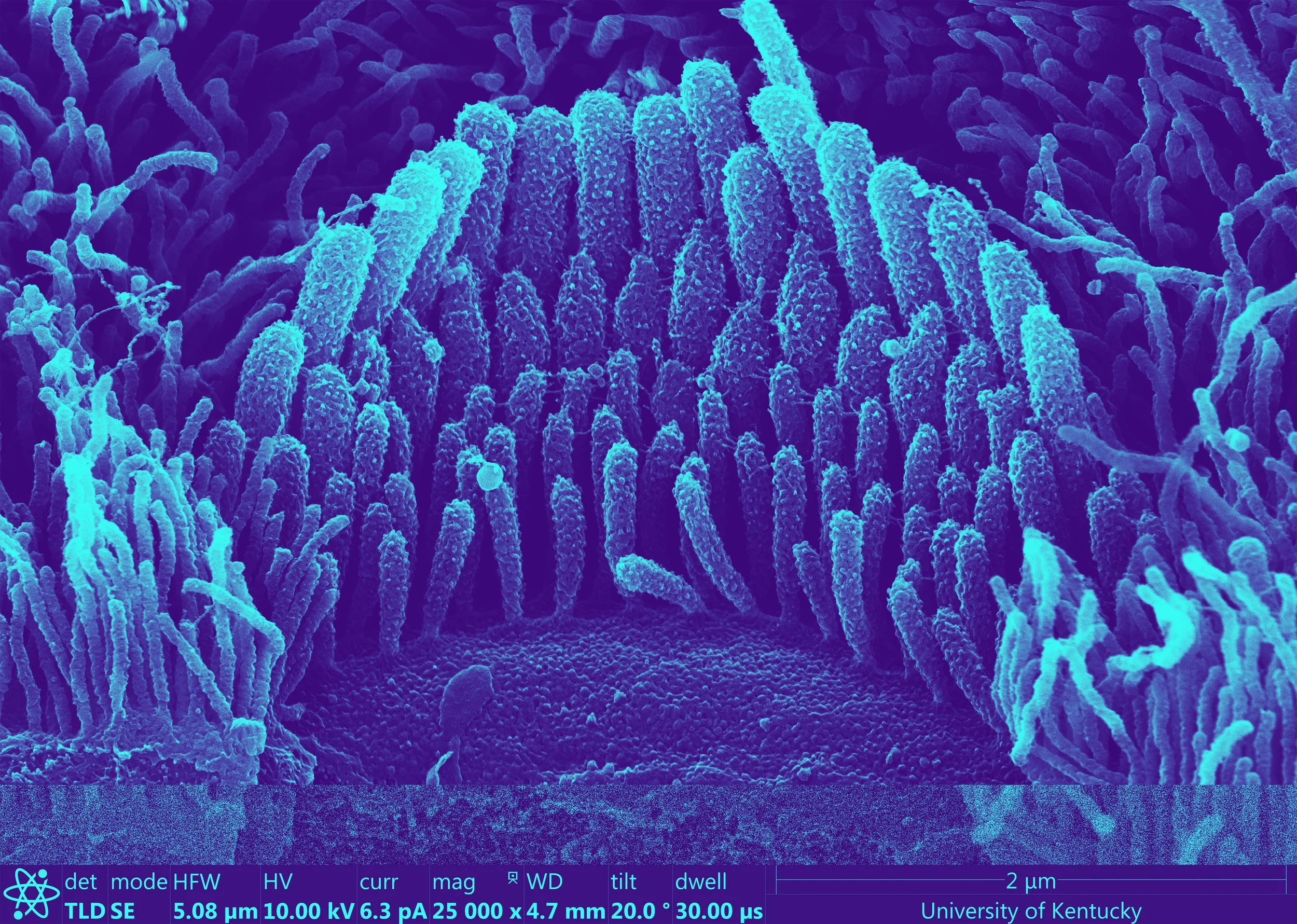
“I'm Lovin' Microscopy” by Abigail Dragich, University of kentucky. imaging facility: EMC (UK). description: Auditory hair cells are the mechanosensitive cells of the cochlea responsible for detecting sound so we can hear. Each cell has a hair bundle structure made of individual stereocilia projections, or “fries”. Image was taken in the UK EMC with scanning electron microscopy on an FEI Helios Nanolab 660.

“Love owl” by Matthew Mulvehill, Conn Center for Renewable Energy Research (uOFL). imaging facility: MNTC huson lab (uofL). description: A scanning electron microscope (SEM) image of the left-over stump of a titanium dioxide nano-rod coated silicon micro-pillar that broke off from the substrate. The dark shadow around the owl’s eyes is silicon, the unique shape being an undesired artifact of a dry reactive ion etch (DRIE) during pillar formation. The silicon is coated with a thin layer of fluorine doped tin oxide followed by the titanium dioxide nano-rods that blanket the entire surface. The micro-pillar that was previously attached to the owl’s face will be used to sustainably generate hydrogen from only water and sunlight.

“micron beach” by Sharmin Akter, university of louisville. imaging facility: mntc huson lab (uofL). Description: This SEM micrograph, captured at the Micro/Nano Technology Center (MNTC) of the University of Louisville, shows a cross-section of a carbon layer deposited on ITO glass. The image was taken to evaluate the uniformity and thickness of the carbon coating. This material serves as a low-cost alternative to conventional metal electrodes in carbon-based perovskite solar cells, improving device scalability and cost-effectiveness.

“The Magic School Bus Gets An Earfull” by Ana Isabel Lopez-Porras, University of kentucky. Co-Author: Evan Magruder. imaging facility: EMC (UK). description: A high resolution scanning electron micrograph showing a mouse inner ear stereocilia bundle. 3D modeled Magic School bus composited over the image using the software Blender.

“When science digs deep, even moles get curious” by Mohammad Junead Mankari, university of louisville. imaging facility: mntc huson lab (UOFL). description: This is a false-colored scanning electron microscope (SEM) image showing the layered microstructure of a material, with different colors representing the elemental distribution of carbon (C), oxygen (O), aluminum (Al), silicon (Si), germanium (Ge), and nitrogen (N).

“nano hulk strength” by Fahad Bin Halim, chemistry department, university of louisville. imaging facility: mntc huson lab (uofl). description: Hydroquinone (HQ)- a monomer is electropolymerized by 15 nm Au nanoparticles (NPs) (Catalyst) to form poly-HQ chains on modified Au/Octanedithiol electrodes. The morphology of such polymeric growth (poly-HQ) shows fractures and splits in its terrain. Applications of poly-HQ include energy storage (Supercapacitor) or energy transfer devices (Fuel cells), Biosensor, Electrochemical sensor. The hammer, lightning and hulk figure was externally added to create a cinematic scene..
Competing in the Most Stunning Category
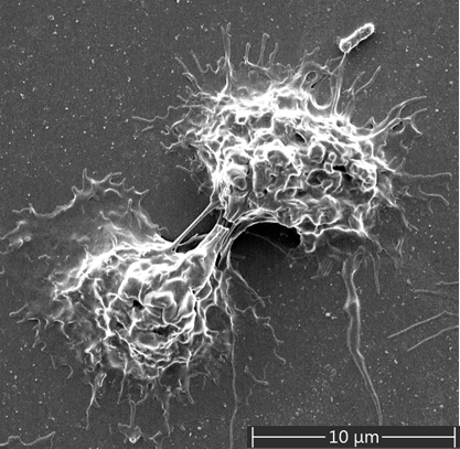
“Guardians in Conversation” by Shikha Srivastava, university of louisville. imaging facility: MNTC huson lab (uofL). Description: Two neutrophils, the body’s first defenders, connect with each other as if quietly communicating during an infection. This small interaction shows how they work together, turning a tiny moment into a powerful display of teamwork and defense.
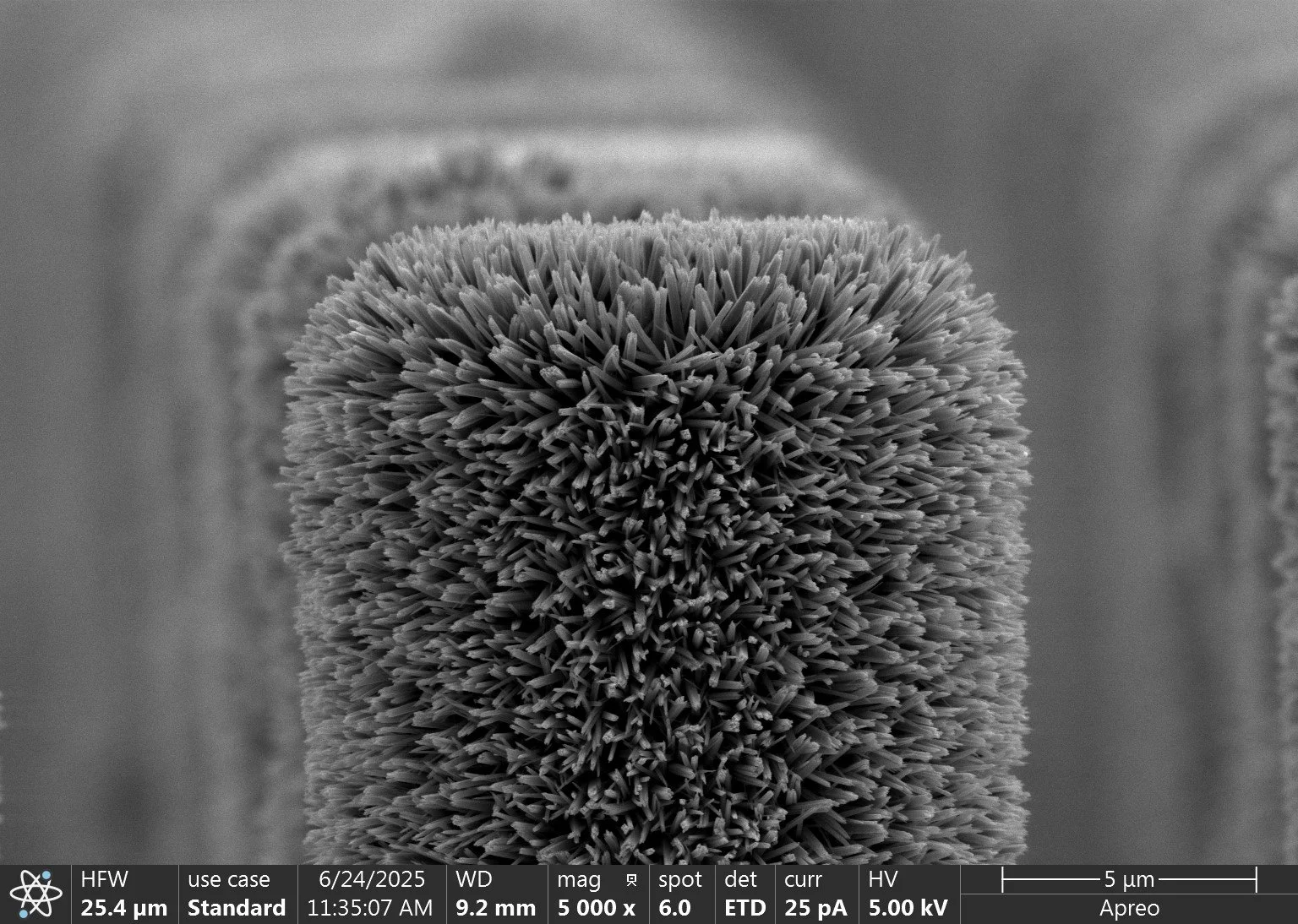
“fuzzy wires” by Matthew Mulvehill, Conn Center for Renewable Energy Research. imaging facility: mntc huson lan (uofL). Description: A scanning electron microscope (SEM) image of the top of a silicon micro-pillar covered with titanium dioxide nano-rods. Both the nano-rods and the underlying silicon micro-pillar can absorb light and use that energy to initiate chemical reactions.

“Anchored signals” by Ana Isabel Lopez-Porras, university of kentucky. imaging facility: emc (uK). description: High-resolution image acquired using Helios Focused Ion Beam-Scanning Electron Microscope (FIB-SEM) system, showing elongated surface projections called stereocilia on the apical surface of an auditory sensory hair cell. The dense actin core of each stereocilium is visible, projecting from the surface of the hair cell. The image also captures the internal organization of the cell, including mitochondria and other organelles beneath the apical membrane.
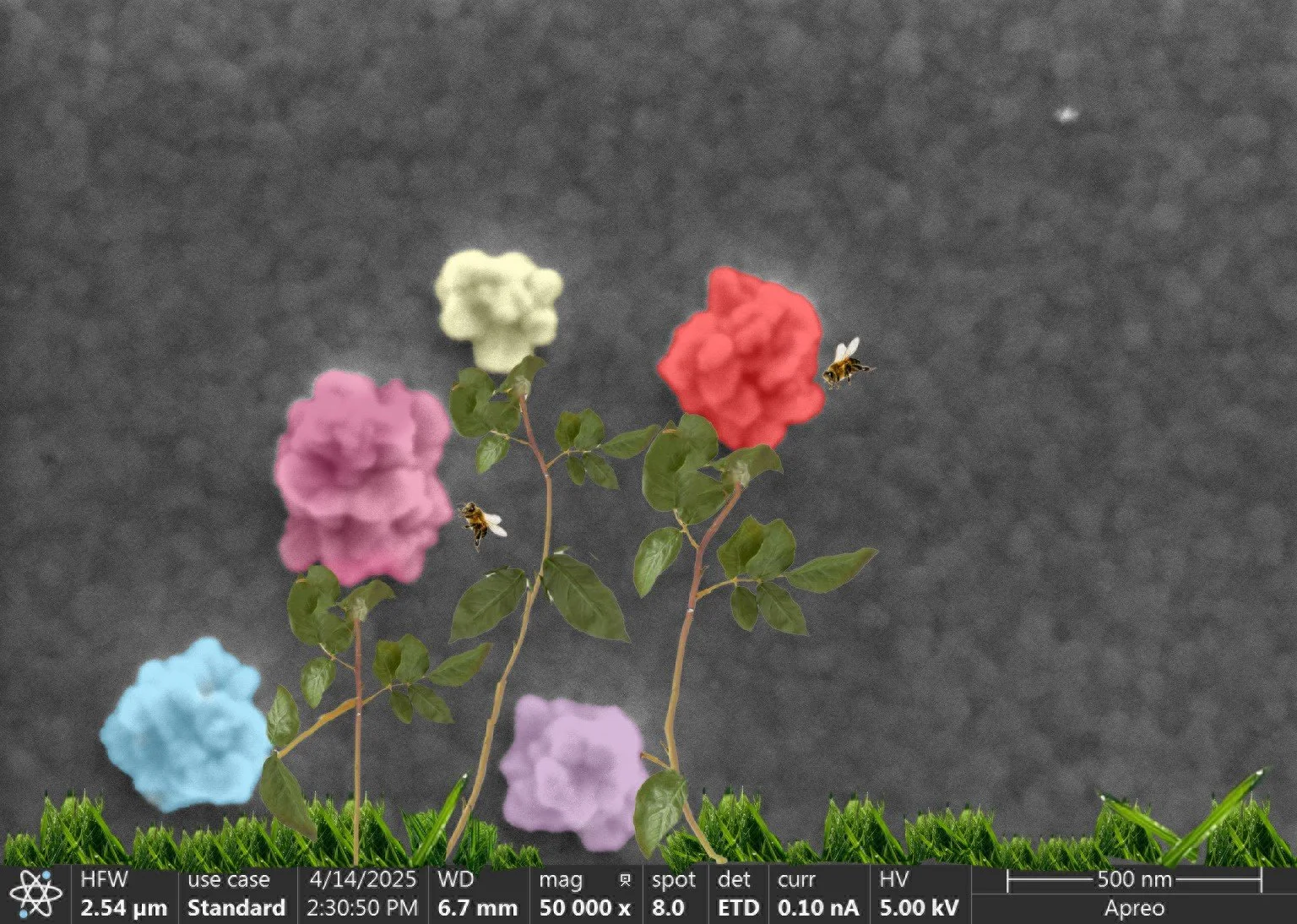
“Bed of nano-roses” by Fahad Bin Halim, chemistry department, university of louisville. imaging facility: mntc huson lab (uofl). description: Tool – Apreo C LoVac FESEM ( Thermo Fisher ) The SEM Image shows how each 4 nm Au nanoparticles (NPs) grow into "Nano-roses" due to electrochemical growth on modified Au/Octanedithiol (ODT) electrodes. Such Au/ODT/Au NP junctions are used in molecular electronic devices ( transistors, gate-switches, break junctions ), sensors, nano-catalyst fabrication. The “Nano-roses” are colorized and leaves, branches, grass, bees are added for viewing pleasure while image details are kept the same.

“Flight Risk” by David Bell. imaging facility: MNTC huson lab (uofl). description:This image shows a cross-section of an emu feather. The sample was prepared by cutting a section of the feather stem and mounting it on a vertical stub. It was then sputter coated with a thin layer of gold to provide a top-down view of the internals of the feather.
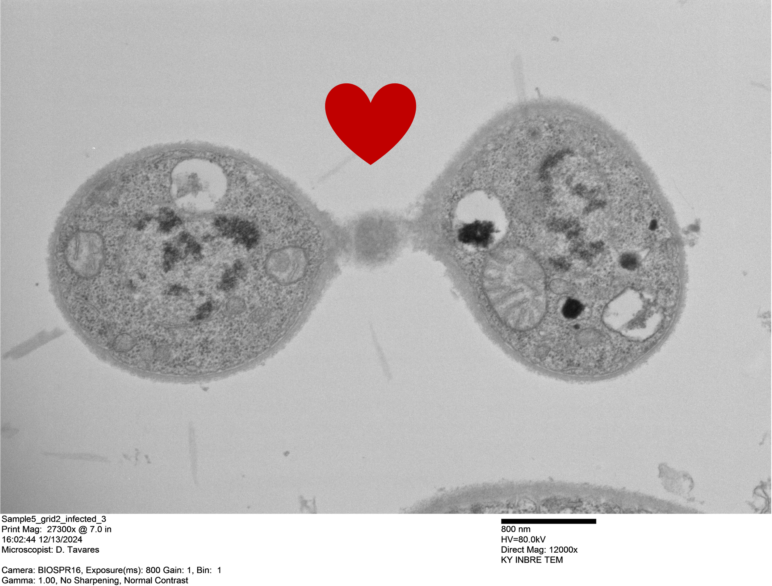
"Love is in the air" by Derica Goncalves Tavares, university of Louisville. co-author: prof. michael perlin. imaging facility: Mntc bioEM Lab (UofL). description: TEM image. UofL MNTC bioEM Lab. Tool: Hitachi HT7700 TEM. Yeast-like cells of the fungal plant pathogen Microbotryum superbum mating, a crucial process that occurs before plant infection

“Co-culture of Lactobacillus crispatus and group B streptococcus” by Nagwa El-Baz. Co-Author: Ryan Doster. imaging faciltiy: mntc Huson lab (uofL). description: Electrospun nanofiber loaded Lactobacillus crispatus (rod-shaped) were applied to the surface of vaginal epithelial cells to treat group B Streptococcus (spherical-shaped) infection. The electrospun nanofibers completely dissolved, releasing live Lactobacillus crispatus.

"battle of the fibers” by Savio Poovathingal, university of kentucky. co-author: Luis Chacon-Olivar. imaging facility: EMC (UK). Description: Comparison of fibers made from rayon and lyocell that are used to manufacture two variants of PICA, a thermal protection system (TPS) material used by NASA on various missions such as Stardust, Mars2020, and Dragonfly.
“Echoes in Blue” by Emma Stevens, university of kentucky. imaging facility: EMC (UK). description: Nanoscale-resolution scanning electron microscopy image of a bundle of stereocilia on top of a single hair cell from the inner ear of the mammalian cochlea. On top of the shortest 2 rows are extracellular tip links, which allow for opening of mechanoelectrical transduction channels to open, and signaling to the brain so that you can hear.

“Lipidless Possibilities” by Payton Nies, University of Kentucky. imaging facility: EMC (UK). Description: Scanning electron microscopy (SEM) image of zoomed in portion of stereocilia hair bundle from the inner ear. These stereocilia have been treated with detergent prior to imaging in the hopes of being able to remove the lipid membrane to see the actin cytoskeleton.
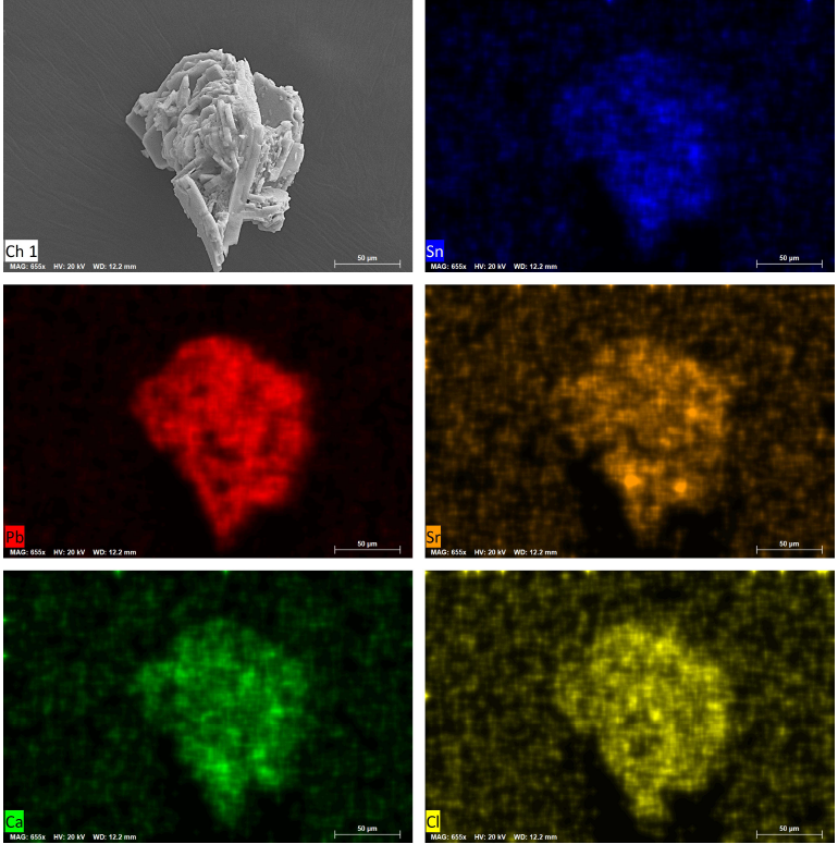
“Nano Firework” by Sharmin Akter, university of louisville. imaging facility: mntc huson lab (uofl). description: SEM image taken at the Micro/Nano Technology Center (MNTC), University of Louisville. SEM image with EDX elemental mapping of a perovskite powder particle, displaying the elemental distribution of Sn, Pb, Sr, Ca, and Cl. The clustered morphology and colorful elemental maps radiate outward like bursts of a firework, inspiring the name Nano Firework. The sample was prepared for use in carbon-based perovskite solar cells